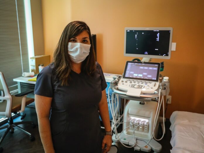
When it comes to women’s reproductive health, few medical advances outshine the diagnostic imaging medical method which is versatile technology to allow physicians to carefully monitor and accurately diagnose health. A special type of imaging test produces information that can be helpful for diagnosing cancer, particularly in soft tissues and it is used as the first step in the standard diagnostic process. Ultrasound Mackay offers many benefits as the test can be performed relatively quickly and cost-effectively without exposing the patient to radiation.This does not produce images with the same level of clarity or detail as tomography and resonance imaging nor can it confirm a cancer diagnosis during a traditional Ultrasound test. A medical professional slowly glides a specialized device or transducer over the patient’s skin in the area of the body being studied as the transducer produces a series of high-frequency sound waves that bounce off the patient’s internal organs compared to X-rays. These are best for taking images of bones as the diagnostic imaging medical method gives a better look at soft tissue means it can do everything from monitoring to detecting potential problems.
The result in the echoes that return to the Ultrasound machine converts the sound waves into two-dimensional images or sonograms that can be viewed in real-time on a monitor as the diagnostic imaging medical method test can also be performed endoscopically. During this minimally invasive imaging procedure, a medical professional inserts an endoscope for a long, flexiblelighted tube with an attached transducer into the patient. Bypassing the transducer over the skin as the shape and intensity of diagnostic imaging medical method echoes can vary depending on the density of the tissue being evaluated. As the sound waves echo differently from fluid-filled cysts and solid masses, a diagnostic imaging medical method can reveal tumors that may be cancerous. As the further testing will be necessary before a cancer diagnosis can be confirmed not only is Ultrasound among the most accurate imaging technologies but they’re also the most cost-effective and accessible. Worrying about the pain or scared to needles which doesn’t have toas thediagnostic imaging medical method is noninvasive and doesn’t require any poking or prodding. Other than a bit of pressure as moving the wand, it shouldn’t feel any discomfort and with tomography scans and X-rays has a risk of exposure to ionising radiation but not with diagnostic imaging medical method.
It uses the power of sound waves to take images of the soft tissues and comes with virtually no harmful effects as the Ultrasound is a staple in prenatal care but it’s also used as a diagnostic tool. If at present with symptoms such as irregular bleeding, cramping, pelvic pain, and bloating, it may indicate an abnormality in the pelvic organs because those symptoms aren’t always easy to diagnose.It is often used thediagnostic imaging medical method to better understand what’s going on and during the diagnostic imaging medical method recline the comfortably on the examination table. Typically, it performed a diagnostic imaging medical method on the outside of the skin as apply a special gel that promotes better sound wave conduction to the treatment area.The part of the body needs to examine as in some cases such as an issue within the ovaries as may order a transvaginal Ultrasound where carefully inserts the wand into the body parts.To get an even closer image of the organs as this type of diagnostic imaging medical method also allows to examine the muscular walls depending on the needs also it may require a sonohysterogram that involves injecting a saline solution and shows if there are any problems with the lining.
It can be unsettling when not sure what’s happening below but pelvic diagnostic imaging medical method givesthe doctor a peek inside to put in mind at ease, whether want to ensure the health or get to the bottom of bothersome symptoms. It is like perplexing cramping or bleeding as the pelvic Ultrasound is a diagnostic test that uses sound waves to create images. These images allow to get a better look at the organs and other tissues in the pelvis as thediagnostic imaging medical method takes place depending on the test’s purposeusing one of two techniques.As part of women’s health care service in transabdominal where it uses an external device on top of the abdomen or transvaginalwhere it is inserted an internal device into the vagina. As the pelvic Ultrasound may be used to assess, monitor or diagnose a range of happenings and conditions and after the diagnostic imaging medical method, it will discuss the results.This includes any additional steps recommendedas the third type of diagnostic imaging medical method a transrectal examis used most commonly to detect prostate problems in men.
However, it may turn to the transrectal Ultrasound for women if a transvaginal exam isn’t feasible or we need to examine the rectum and during the procedure, it will insert a probe into the rectum that sends sound waves to internal tissues. During a transabdominal pelvic diagnostic imaging it will feel a cool, wet sensation when applying a lubricating gel to abdomen or pelvic region. A transvaginal pelvic diagnostic imaging is a bit more invasive and the process isn’t usually painful although it may notice added tenderness if already experiencing the pain. It may notice a sense of fullness or pressure during the test but taking special care to keep at ease as the transrectal diagnostic imaging which uses a cool gel for lubrication.It may cause some mild discomfort which can minimise by lying still as the main reason ofUltrasound is preferred over X-ray and it’s safer for pregnant women and their unborn children. Thediagnostic imaging has other advantages as well rather than radiation as thediagnostic imaging generates images by sending soundwaves through the skin and into internal tissues as these diagnostic imaging is noninvasive and painless but it’s also much safer.


